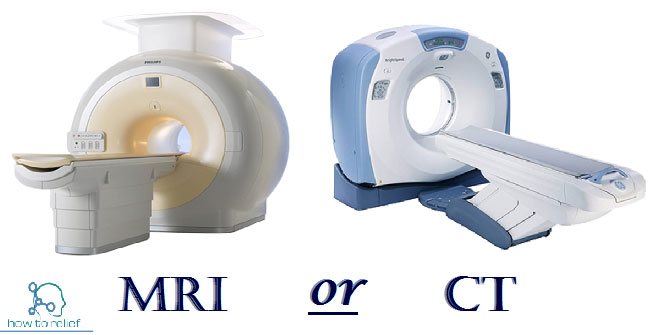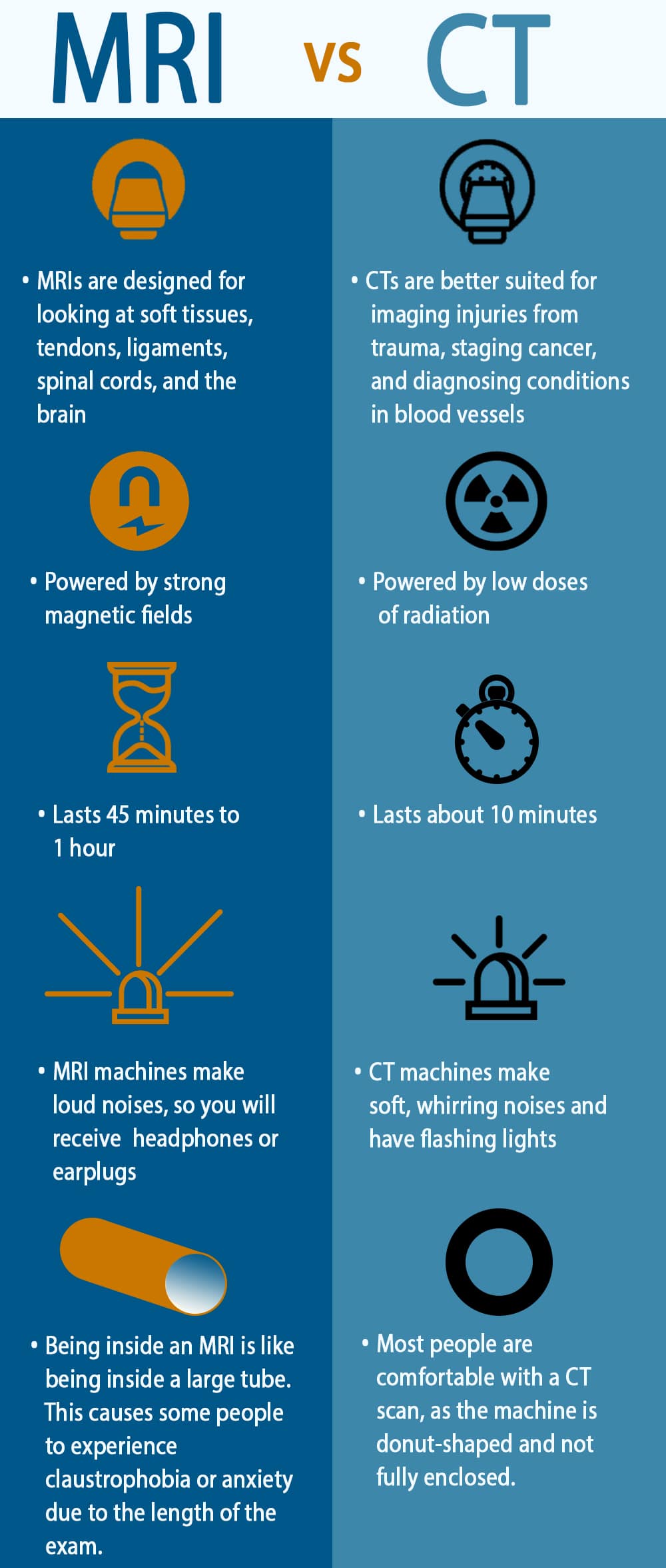

MRI images, on the other hand, use a strong magnetic field, which can attract metal objects or may cause metal in your body to move. For example, the radiation exposure from one low-dose CT scan of the chest is the same amount of radiation as every person receives from the earth’s natural background radiation over six months. This exposure to radiation in a CT scan is higher than that of standard X-rays, although the associated risk is still small. ARE THESE EXAMS SAFE?Īn important difference between CT and MRI is that a CT scan involves radiation. Whether it’s identifying a targeted area for treatment with surgery or an injection, finding an illness early, or excluding serious pathology, a clear diagnosis can provide peace of mind for patients. A CT may be preferred to evaluate fractures or look for cancers or blood clots. CT scans, in comparison, are best for imaging bone, soft tissues in the chest or abdomen, and blood vessels. For example, MRI is very good at examining soft tissues such as tendons and ligaments, evaluating the spinal cord, and identifying strokes in the brain. Whether a doctor requests a CT or an MRI scan often depends on the presumed diagnosis. This data is transmitted to a computer, which builds a 3D cross-sectional picture of area the body being scanned. CT can demonstrate different levels of tissue density.

#CT AND MRI DIAGNOSTICS SERIES#
While a standard X-ray machine sends only one radiation beam, a CT scanner emits a series of beams as it moves in an arc around the body. MRI images can be taken of most body parts.Ī CT scan is made from X-rays. A radio frequency pulse is then directed at a specific area of the body, while smaller magnets are used to alter the magnetic field on a small, but localized level.Īs tissues responds differently to these magnetic field alterations, a computer can convert the data into a picture. HOW DO THEY WORK?Īn MRI scan creates images by exposing hydrogen atoms within our body to a magnetic field which controls the direction and frequency at which these hydrogen protons spin. This may influence which modality is best for a specific diagnosis,” says Rick Myszkowski, lead MRI technologist and Mayfair Place clinic manager. “Although both CT and MRI are excellent tools for diagnostic imaging, each has its own strengths. A patient’s medical and family history, risk factors, and type and duration of symptoms, all affect the physician’s decision on which type of imaging is appropriate. They are often ordered when more detail is needed, or the cause of symptoms is unclear during a physical exam or on other types of imaging.īut sometimes it can be confusing to understand why one exam is requested and not the other, or why a patient might be sent for both types of scans. Computed tomography (CT) and magnetic resonance imaging (MRI) are two types of medical imaging that have a reputation for providing a thorough look inside the body.


 0 kommentar(er)
